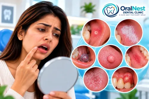
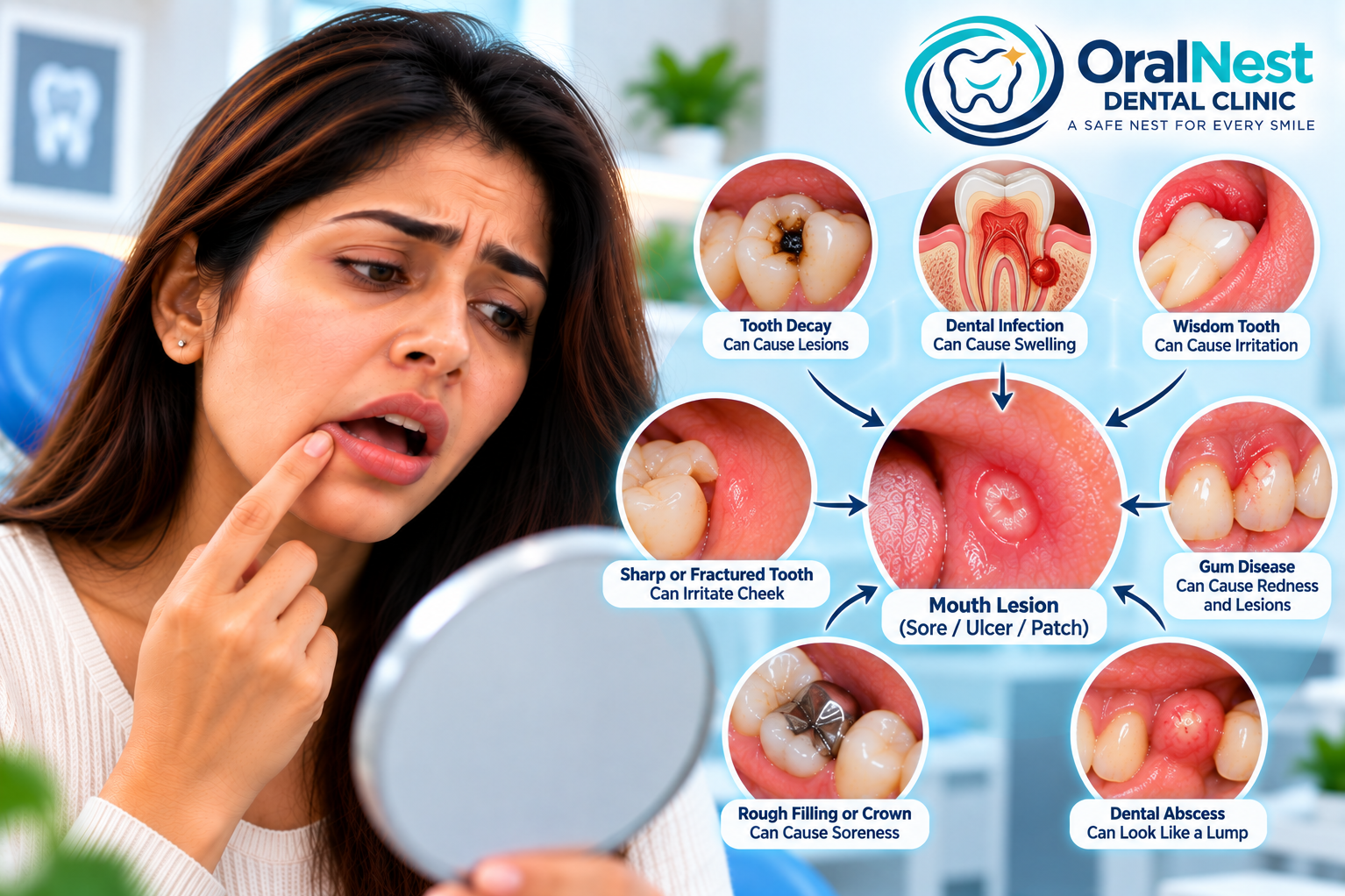
A sore, swelling, ulcer, red area or unusual patch inside the mouth may appear to be a problem of the oral lining itself.
But sometimes, the real source is a tooth or the tissues surrounding it.
This is one reason oral diagnosis can be more complicated than simply looking at the visible lesion.
A dental infection, fractured tooth, erupting wisdom tooth, periodontal problem or irritation from a restoration can sometimes produce changes in nearby soft tissues.
So when you notice a lesion, the important question is not only:
“What does this area look like?”
It is also:
“Could something underneath or beside it be causing this change?”
How can a tooth create a mouth lesion?
Teeth and the surrounding oral tissues are closely connected.
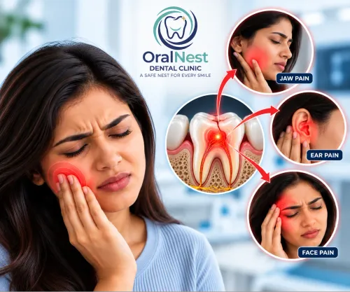
When a tooth develops decay, infection, fracture or another problem, inflammation can sometimes extend into the surrounding tissues.
This may produce:
swelling
tenderness
redness
a draining sinus or gum “pimple”
localized soreness
recurrent swelling
changes in nearby soft tissue
A patient may notice the visible change in the gum or cheek and assume that the mucosa itself is the primary problem.
Sometimes the actual source is the tooth.
Can a dental infection look like a gum swelling?
Yes.
A tooth with an infection around its root may sometimes produce a localized swelling or draining point on the gum.
The tooth itself may not always be severely painful.
This can lead to a common misunderstanding:
“There is a bump on my gum, so I just need something for the gum.”
But if the swelling is being caused by an infected tooth, treating only the visible swelling does not address the source.
The tooth and surrounding tissues need to be evaluated together.
NIDCR notes that an infected tooth can lead to an abscess, which may cause pain, swelling and other symptoms.
What does a dental abscess look like?
A dental abscess can present in different ways.
Some patients experience:
significant toothache
swelling
tenderness
sensitivity
facial swelling
fever
Others may notice a smaller recurring swelling on the gum that occasionally drains.
The appearance can therefore vary.
This is why a gum swelling should not be diagnosed simply by looking at it.
The clinician may need to determine whether the source is:
the tooth → the supporting tissues → the gum → or the oral mucosa itself.
Can a wisdom tooth create a mouth lesion?
Yes.
A partially erupted or impacted wisdom tooth can contribute to inflammation of the surrounding gum.
The tissue around the tooth may become swollen, tender or irritated.
Food accumulation and difficulty cleaning the area can further contribute to local inflammation.
Sometimes the cheek may also become irritated by the position of the tooth.
However, not every lesion near the back of the mouth is caused by a wisdom tooth.
The position of the tooth and the condition of the surrounding tissues need to be assessed.
Can a sharp tooth create a white or red patch?
Yes.
Repeated mechanical irritation from a sharp tooth, fractured tooth or rough restoration can affect the adjacent oral lining.
The tissue may become:
sore
red
thickened
ulcerated
or develop a whitish appearance
This is an important example of why identifying the source of irritation matters.
If the cause is not removed, repeatedly applying medication to the lesion may not solve the underlying problem.
Can a dental filling or crown irritate the cheek?
It can.
An irregular restoration, altered contour or rough edge may repeatedly contact the cheek, tongue or gum.
The resulting irritation may appear exactly where the restoration contacts the tissue.
A patient may notice:
“This sore keeps coming back in the same place.”
That recurring location can be a useful clue.
During examination, the clinician may assess the restoration, tooth shape, bite and relationship between the dental structure and the lesion.
Can gum disease look like a mouth lesion?
Gum inflammation can produce redness, swelling, tenderness and bleeding around the teeth.
The changes may sometimes be mistaken for a generalized oral mucosal problem.
NIDCR describes gum disease as potentially causing red, swollen or tender gums and bleeding.
The distinction matters because the treatment approach depends on whether the problem originates primarily from:
plaque and gum inflammation
periodontal tissues
a specific tooth
oral mucosa
or another underlying condition
A proper examination helps separate these possibilities.
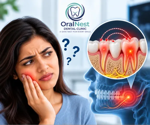
Can a dental infection cause a lesion somewhere nearby?
Yes.
The visible swelling does not always appear exactly where a patient expects the problem to be.
The pathway of infection and inflammation can influence where the swelling becomes noticeable.
This is particularly important when evaluating:
upper teeth and nearby tissues
lower molars
wisdom teeth
facial swelling
recurrent gum swellings
draining points
The clinician may therefore examine both the lesion and nearby teeth rather than treating the visible area in isolation.
What if the lesion is on the cheek next to a tooth?
This is a classic situation where the source may not be immediately obvious.
The cheek could be affected by:
repeated biting
a sharp tooth
a fractured tooth
a rough restoration
an erupting tooth
an oral mucosal condition
The location of the lesion relative to the teeth can provide important information.
For example, if a lesion occurs exactly where a sharp tooth repeatedly contacts the cheek, mechanical irritation becomes an important consideration.
But if there is no obvious source of trauma, the lesion itself may require closer evaluation.
Can a dental problem look like an ulcer?
Sometimes.
A traumatic ulcer caused by a sharp tooth can look like a conventional mouth ulcer.
The patient may therefore repeatedly apply an ulcer gel while the tooth continues to traumatize the tissue.
This creates an important diagnostic principle:
If an ulcer repeatedly occurs in exactly the same location, look for a repeated local cause.
That could include a tooth, restoration, appliance or biting habit.
If the lesion persists after the suspected source is addressed, the diagnosis should be reconsidered.
Can a dental problem look like a lump?
Yes.
A swelling caused by a dental infection may appear as a small lump or pimple-like structure on the gum.
Likewise, inflammation around an erupting tooth can produce localized swelling.
But oral lumps can also arise from many non-dental causes.
Therefore, the presence of a lump near a tooth does not automatically prove that the tooth is responsible.
The clinical relationship between the two needs to be established.
What if the tooth doesn't hurt?
This is an important point.
A dental source can sometimes exist without dramatic tooth pain.
For example, an infection may drain through surrounding tissues and reduce pressure, changing how the patient experiences symptoms.
Therefore:
No toothache does not automatically mean no dental problem.
A persistent swelling, draining point or unexplained lesion near a tooth may still justify examination of that tooth.
Can a lesion be caused by something other than the teeth?
Absolutely.
This is why diagnosis must remain open.
An oral lesion may be related to:
trauma
infection
salivary gland problems
inflammatory conditions
oral mucosal disorders
medication effects
systemic factors
or other causes
The purpose of a diagnosis-focused examination is not to prove that every lesion is dental.
It is to determine whether the teeth are involved or not.
How do you determine whether the tooth is responsible?
The evaluation may involve several steps.
1. Clinical examination
The tooth, gums and surrounding lesion are examined carefully.
2. Symptom history
The clinician may ask when the lesion appeared, whether it recurs and whether there is pain or sensitivity.
3. Tooth assessment
The suspected tooth may be evaluated for decay, fracture, previous treatment, periodontal problems or other abnormalities.
4. Radiographic assessment
When indicated, dental imaging may help evaluate structures that cannot be seen directly.
5. Examination of the lesion itself
The colour, surface, borders, texture, size and behaviour of the oral lesion are considered.
The aim is to connect the findings rather than treating each finding separately.
Why does the correct diagnosis change the treatment?
Consider two patients who both have a small swelling on the gum.
Patient A
The swelling is related to an infected tooth.
The appropriate treatment needs to address the dental source.
Patient B
The swelling is unrelated to the tooth and represents a separate oral soft-tissue condition.
Treating the tooth would not necessarily solve the problem.
This simple comparison explains why diagnosis must come before treatment.
The same visible symptom can lead to completely different treatment decisions depending on its cause.
What if the lesion remains after dental treatment?
This is an important clinical situation.
If a suspected dental cause has been appropriately addressed but the lesion does not resolve as expected, the diagnosis should be reassessed.
The original assumption may have been incomplete.
Further examination may then be appropriate to determine whether:
another source of irritation remains
the lesion requires additional observation
another dental problem exists
the lesion is primarily mucosal
further investigation is needed
A good diagnosis is not simply the first explanation that seems possible.
It is the explanation that continues to fit the clinical evidence.
When should a mouth lesion be checked?
Consider evaluation when a lesion:
persists longer than expected
repeatedly returns
appears beside a problematic tooth
is associated with swelling
bleeds
develops a red or white colour change
becomes firm
causes persistent pain
produces numbness
is associated with difficulty eating or swallowing
has no obvious explanation
NIDCR recommends evaluation of persistent oral sores, lumps, thickened areas, and red or white patches, particularly when they last more than two weeks.
That recommendation is about evaluation, not about assuming that the lesion represents cancer.
The visible lesion may not be the real problem
This is one of the most important concepts in oral diagnosis.
A patient sees:
a sore
But the cause may be:
a sharp tooth.
A patient sees:
a gum swelling
But the source may be:
an infected tooth.
A patient sees:
a cheek lesion
But the cause may be:
repeated trauma from the bite.
And sometimes the visible lesion truly is a condition of the oral mucosa itself.
The challenge is determining which explanation fits the evidence.
Oral Medicine & Oral Diagnosis at OralNest Dental Clinic | Wakad
At OralNest Dental Clinic | Wakad, an unexplained oral lesion is not evaluated only as a surface problem.
The examination considers the lesion, neighbouring teeth, gums, bite, possible sources of trauma, salivary structures and relevant clinical history to determine whether the visible finding has a dental source or requires a broader oral diagnosis.
The objective is simple:
Find the source before deciding on the treatment.
For patients from Wakad and nearby areas including Hinjawadi, Tathawade, Punawale, Pimple Saudagar, Pimple Gurav and Rahatani, persistent or unexplained oral lesions can be evaluated through a structured oral diagnosis.
Clinic Timings
Monday–Saturday: 5:30 PM–9:00 PM
Sunday: 10:00 AM–1:00 PM
Prior appointment is convenient.
At OralNest Dental Clinic | Wakad
Understand First. Diagnose Carefully. Decide With Clarity.
But sometimes, the real source is a tooth or the tissues surrounding it.
This is one reason oral diagnosis can be more complicated than simply looking at the visible lesion.
A dental infection, fractured tooth, erupting wisdom tooth, periodontal problem or irritation from a restoration can sometimes produce changes in nearby soft tissues.
So when you notice a lesion, the important question is not only:
“What does this area look like?”
It is also:
“Could something underneath or beside it be causing this change?”
How can a tooth create a mouth lesion?
Teeth and the surrounding oral tissues are closely connected.
When a tooth develops decay, infection, fracture or another problem, inflammation can sometimes extend into the surrounding tissues.
This may produce:
swelling
tenderness
redness
a draining sinus or gum “pimple”
localized soreness
recurrent swelling
changes in nearby soft tissue
A patient may notice the visible change in the gum or cheek and assume that the mucosa itself is the primary problem.
Sometimes the actual source is the tooth.
Can a dental infection look like a gum swelling?
Yes.
A tooth with an infection around its root may sometimes produce a localized swelling or draining point on the gum.
The tooth itself may not always be severely painful.
This can lead to a common misunderstanding:
“There is a bump on my gum, so I just need something for the gum.”
But if the swelling is being caused by an infected tooth, treating only the visible swelling does not address the source.
The tooth and surrounding tissues need to be evaluated together.
NIDCR notes that an infected tooth can lead to an abscess, which may cause pain, swelling and other symptoms.
What does a dental abscess look like?
A dental abscess can present in different ways.
Some patients experience:
significant toothache
swelling
tenderness
sensitivity
facial swelling
fever
Others may notice a smaller recurring swelling on the gum that occasionally drains.
The appearance can therefore vary.
This is why a gum swelling should not be diagnosed simply by looking at it.
The clinician may need to determine whether the source is:
the tooth → the supporting tissues → the gum → or the oral mucosa itself.
Can a wisdom tooth create a mouth lesion?
Yes.
A partially erupted or impacted wisdom tooth can contribute to inflammation of the surrounding gum.
The tissue around the tooth may become swollen, tender or irritated.
Food accumulation and difficulty cleaning the area can further contribute to local inflammation.
Sometimes the cheek may also become irritated by the position of the tooth.
However, not every lesion near the back of the mouth is caused by a wisdom tooth.
The position of the tooth and the condition of the surrounding tissues need to be assessed.
Can a sharp tooth create a white or red patch?
Yes.
Repeated mechanical irritation from a sharp tooth, fractured tooth or rough restoration can affect the adjacent oral lining.
The tissue may become:
sore
red
thickened
ulcerated
or develop a whitish appearance
This is an important example of why identifying the source of irritation matters.
If the cause is not removed, repeatedly applying medication to the lesion may not solve the underlying problem.
Can a dental filling or crown irritate the cheek?
It can.
An irregular restoration, altered contour or rough edge may repeatedly contact the cheek, tongue or gum.
The resulting irritation may appear exactly where the restoration contacts the tissue.
A patient may notice:
“This sore keeps coming back in the same place.”
That recurring location can be a useful clue.
During examination, the clinician may assess the restoration, tooth shape, bite and relationship between the dental structure and the lesion.
Can gum disease look like a mouth lesion?
Gum inflammation can produce redness, swelling, tenderness and bleeding around the teeth.
The changes may sometimes be mistaken for a generalized oral mucosal problem.
NIDCR describes gum disease as potentially causing red, swollen or tender gums and bleeding.
The distinction matters because the treatment approach depends on whether the problem originates primarily from:
plaque and gum inflammation
periodontal tissues
a specific tooth
oral mucosa
or another underlying condition
A proper examination helps separate these possibilities.
Can a dental infection cause a lesion somewhere nearby?
Yes.
The visible swelling does not always appear exactly where a patient expects the problem to be.
The pathway of infection and inflammation can influence where the swelling becomes noticeable.
This is particularly important when evaluating:
upper teeth and nearby tissues
lower molars
wisdom teeth
facial swelling
recurrent gum swellings
draining points
The clinician may therefore examine both the lesion and nearby teeth rather than treating the visible area in isolation.
What if the lesion is on the cheek next to a tooth?
This is a classic situation where the source may not be immediately obvious.
The cheek could be affected by:
repeated biting
a sharp tooth
a fractured tooth
a rough restoration
an erupting tooth
an oral mucosal condition
The location of the lesion relative to the teeth can provide important information.
For example, if a lesion occurs exactly where a sharp tooth repeatedly contacts the cheek, mechanical irritation becomes an important consideration.
But if there is no obvious source of trauma, the lesion itself may require closer evaluation.
Can a dental problem look like an ulcer?
Sometimes.
A traumatic ulcer caused by a sharp tooth can look like a conventional mouth ulcer.
The patient may therefore repeatedly apply an ulcer gel while the tooth continues to traumatize the tissue.
This creates an important diagnostic principle:
If an ulcer repeatedly occurs in exactly the same location, look for a repeated local cause.
That could include a tooth, restoration, appliance or biting habit.
If the lesion persists after the suspected source is addressed, the diagnosis should be reconsidered.
Can a dental problem look like a lump?
Yes.
A swelling caused by a dental infection may appear as a small lump or pimple-like structure on the gum.
Likewise, inflammation around an erupting tooth can produce localized swelling.
But oral lumps can also arise from many non-dental causes.
Therefore, the presence of a lump near a tooth does not automatically prove that the tooth is responsible.
The clinical relationship between the two needs to be established.
What if the tooth doesn't hurt?
This is an important point.
A dental source can sometimes exist without dramatic tooth pain.
For example, an infection may drain through surrounding tissues and reduce pressure, changing how the patient experiences symptoms.
Therefore:
No toothache does not automatically mean no dental problem.
A persistent swelling, draining point or unexplained lesion near a tooth may still justify examination of that tooth.
Can a lesion be caused by something other than the teeth?
Absolutely.
This is why diagnosis must remain open.
An oral lesion may be related to:
trauma
infection
salivary gland problems
inflammatory conditions
oral mucosal disorders
medication effects
systemic factors
or other causes
The purpose of a diagnosis-focused examination is not to prove that every lesion is dental.
It is to determine whether the teeth are involved or not.
How do you determine whether the tooth is responsible?
The evaluation may involve several steps.
1. Clinical examination
The tooth, gums and surrounding lesion are examined carefully.
2. Symptom history
The clinician may ask when the lesion appeared, whether it recurs and whether there is pain or sensitivity.
3. Tooth assessment
The suspected tooth may be evaluated for decay, fracture, previous treatment, periodontal problems or other abnormalities.
4. Radiographic assessment
When indicated, dental imaging may help evaluate structures that cannot be seen directly.
5. Examination of the lesion itself
The colour, surface, borders, texture, size and behaviour of the oral lesion are considered.
The aim is to connect the findings rather than treating each finding separately.
Why does the correct diagnosis change the treatment?
Consider two patients who both have a small swelling on the gum.
Patient A
The swelling is related to an infected tooth.
The appropriate treatment needs to address the dental source.
Patient B
The swelling is unrelated to the tooth and represents a separate oral soft-tissue condition.
Treating the tooth would not necessarily solve the problem.
This simple comparison explains why diagnosis must come before treatment.
The same visible symptom can lead to completely different treatment decisions depending on its cause.
What if the lesion remains after dental treatment?
This is an important clinical situation.
If a suspected dental cause has been appropriately addressed but the lesion does not resolve as expected, the diagnosis should be reassessed.
The original assumption may have been incomplete.
Further examination may then be appropriate to determine whether:
another source of irritation remains
the lesion requires additional observation
another dental problem exists
the lesion is primarily mucosal
further investigation is needed
A good diagnosis is not simply the first explanation that seems possible.
It is the explanation that continues to fit the clinical evidence.
When should a mouth lesion be checked?
Consider evaluation when a lesion:
persists longer than expected
repeatedly returns
appears beside a problematic tooth
is associated with swelling
bleeds
develops a red or white colour change
becomes firm
causes persistent pain
produces numbness
is associated with difficulty eating or swallowing
has no obvious explanation
NIDCR recommends evaluation of persistent oral sores, lumps, thickened areas, and red or white patches, particularly when they last more than two weeks.
That recommendation is about evaluation, not about assuming that the lesion represents cancer.
The visible lesion may not be the real problem
This is one of the most important concepts in oral diagnosis.
A patient sees:
a sore
But the cause may be:
a sharp tooth.
A patient sees:
a gum swelling
But the source may be:
an infected tooth.
A patient sees:
a cheek lesion
But the cause may be:
repeated trauma from the bite.
And sometimes the visible lesion truly is a condition of the oral mucosa itself.
The challenge is determining which explanation fits the evidence.
Oral Medicine & Oral Diagnosis at OralNest Dental Clinic | Wakad
At OralNest Dental Clinic | Wakad, an unexplained oral lesion is not evaluated only as a surface problem.
The examination considers the lesion, neighbouring teeth, gums, bite, possible sources of trauma, salivary structures and relevant clinical history to determine whether the visible finding has a dental source or requires a broader oral diagnosis.
The objective is simple:
Find the source before deciding on the treatment.
For patients from Wakad and nearby areas including Hinjawadi, Tathawade, Punawale, Pimple Saudagar, Pimple Gurav and Rahatani, persistent or unexplained oral lesions can be evaluated through a structured oral diagnosis.
Clinic Timings
Monday–Saturday: 5:30 PM–9:00 PM
Sunday: 10:00 AM–1:00 PM
Prior appointment is convenient.
At OralNest Dental Clinic | Wakad
Understand First. Diagnose Carefully. Decide With Clarity.
Keywords