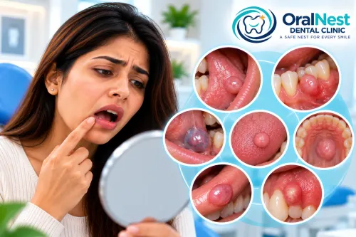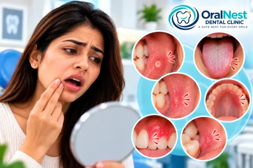
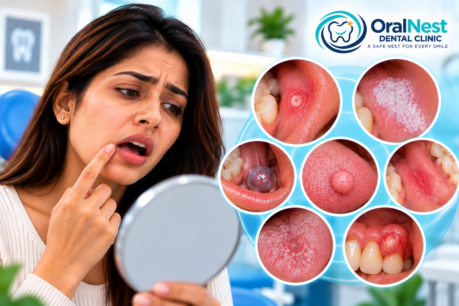
Finding a sore, patch, ulcer, lump or unusual area inside the mouth does not automatically mean that something serious is wrong.
Many oral lesions have simple explanations such as trauma, irritation, infection or inflammation.
But sometimes a lesion cannot be confidently explained by its appearance alone.
That is when further investigation may become important.
The purpose is not to create fear.
It is to answer a more useful question:
“What exactly is this change in the oral tissue?”
A careful diagnosis can prevent both unnecessary treatment and delayed attention to a lesion that genuinely needs investigation.
What does “further investigation” actually mean?
Patients often hear the phrase and immediately think of a biopsy.
But further investigation does not always mean biopsy.
Depending on the clinical situation, it may simply involve:
a more detailed oral examination
documenting the lesion and monitoring it
checking for a source of trauma
examining nearby teeth
reviewing medications and medical history
assessing tobacco or other habits
appropriate imaging
laboratory investigations when relevant
referral to another specialist
or, in selected cases, tissue sampling
The investigation is chosen according to the clinical question that needs to be answered.
Why can't every oral lesion be diagnosed just by looking?
Because different conditions can sometimes have overlapping appearances.
A white patch may represent surface coating, irritation, infection or a mucosal condition.
A red area may be inflammatory, traumatic or something that requires closer assessment.
A lump may arise from trauma, a tooth, salivary tissue, a cyst or another soft-tissue process.
Therefore:
Appearance provides clues. It does not always provide the diagnosis.
This is why oral diagnosis involves history, examination and clinical reasoning rather than simply matching a lesion to a photograph.
When does a lesion become more important to investigate?
Several features may increase the need for closer assessment.
These can include a lesion that:
persists without an obvious explanation
does not heal as expected
repeatedly returns
increases in size
changes in colour
develops a different surface texture
becomes firm or indurated
bleeds without an obvious cause
is associated with persistent pain
produces numbness
occurs as a persistent red or white patch
is associated with difficulty chewing or swallowing
has no identifiable source of irritation
NIDCR advises that persistent sores, lumps, thick patches, and white or red patches lasting more than two weeks should be evaluated by a dentist or doctor.
This does not mean that a lesion present for two weeks is automatically dangerous.
It means persistence is an important reason to stop assuming and start evaluating.
What if the lesion does not hurt?
A painless lesion can still require assessment.
Pain is only one characteristic.
Some oral conditions produce significant discomfort.
Others may remain completely symptom-free and only become noticeable when a patient looks in the mirror or when a clinician performs an examination.
Therefore:
“It doesn't hurt” is not the same as “it doesn't need attention.”
The duration, appearance, location, texture and changes over time can be equally important.
What if the lesion looks small?
Size alone does not determine significance.
A small lesion can be completely harmless.
A small persistent lesion can also deserve evaluation if its cause is unclear.
Similarly, a larger lesion may have a straightforward explanation such as trauma or inflammation.
This is why clinicians do not generally make decisions based on size alone.
What if I think I know what caused it?
This is common.
A patient may think:
“I must have bitten myself.”
Or:
“It is probably a fungal infection.”
Or:
“It must be because of my wisdom tooth.”
Sometimes the assumption is correct.
But if the lesion does not behave as expected after the suspected cause is removed or treated, the original explanation should be reconsidered.
A diagnosis should remain open to revision when the clinical course does not fit.
Why is persistence such an important clue?
Healthy oral tissues can experience minor injuries and irritation, and many of these changes resolve as the tissue heals.
When an area remains unchanged, repeatedly returns or progressively changes, the clinical question becomes different.
Instead of:
“How do I make this sore feel better?”
the question becomes:
“Why isn't this tissue returning to normal?”
That shift—from symptom relief to cause identification—is an important part of diagnosis-focused oral care.
Does a red or white patch always need a biopsy?
No.
A biopsy is not automatically required for every red or white oral lesion.
Some lesions can be confidently explained clinically.
Others may be observed after removing an obvious source of irritation.
If a lesion remains unexplained or has features that warrant further assessment, tissue sampling may be considered.
The decision depends on the clinical findings and the level of diagnostic certainty.
NIDCR notes that when an oral examination identifies something unusual, additional testing or referral may be recommended.
What is the purpose of a biopsy?
A biopsy involves taking a small sample of tissue for microscopic examination.
Its purpose is to answer a diagnostic question that cannot always be answered through visual examination alone.
A biopsy can help determine what type of tissue change is present and whether further treatment or monitoring is necessary.
Importantly:
Being advised to have a biopsy does not mean that cancer has been diagnosed.
It means that obtaining tissue information may provide a more definitive answer.
Why early evaluation matters
The mouth is an area where changes can sometimes be seen and examined directly.
This makes regular oral examination valuable.
The American Academy of Oral Medicine states that risk-factor assessment and visual and tactile examination of the oral mucosa are part of routine oral health evaluation.
When a persistent abnormality is identified, understanding it earlier can help determine the appropriate next step rather than allowing uncertainty to continue.
Does further investigation mean something serious is suspected?
Not necessarily.
This is an important distinction.
Further investigation can simply mean:
“The current information is not enough to confidently explain this finding.”
For example, a lesion may look unusual but still have a benign explanation.
The clinician may investigate because it is better to establish the cause than to guess.
This approach protects patients from two opposite problems:
Unnecessary alarm
Assuming every unusual lesion is dangerous.
False reassurance
Assuming every persistent lesion is harmless.
Good diagnosis aims for neither.
What happens during a detailed oral examination?
The assessment usually begins with the patient's history.
Important questions may include:
When was the lesion first noticed?
Has it changed?
Has it increased in size?
Does it come and go?
Is there pain, burning or bleeding?
Was there any trauma?
Is there a sharp tooth nearby?
Are dentures or appliances contacting it?
Are there tobacco or other relevant habits?
What medications are being taken?
Have similar lesions appeared previously?
The clinician then examines the entire oral cavity rather than focusing only on the area the patient noticed.
This broader examination can sometimes reveal additional information that changes the initial impression.
Why examining the whole mouth matters
An oral lesion should not always be considered in isolation.
The surrounding tissue, other parts of the mouth, teeth, gums, tongue and salivary structures may provide useful clues.
A patient may come because of one white patch, for example, but the examination may identify dryness, repeated trauma or another relevant finding elsewhere.
This is why a comprehensive oral examination can be more informative than simply examining the visible spot.
Can imaging help?
In selected situations, yes.
Imaging may be useful when the lesion is associated with:
a tooth
bone
an impacted tooth
a swelling
a suspected cyst
deeper structures that cannot be evaluated adequately by surface examination
But imaging is not automatically required for every mucosal lesion.
The appropriate investigation depends on the question being asked.
The test should follow the clinical question—not the other way around.
What if the lesion disappears before the appointment?
That information is still useful.
If possible, note:
when it appeared
how long it lasted
where it occurred
what it looked like
whether it caused pain
whether anything seemed to trigger it
A photograph taken during the episode may also help provide a record, although a photograph cannot replace clinical examination.
For recurring lesions, documenting the pattern can help distinguish temporary changes from persistent or recurrent conditions.
Why self-treatment can sometimes complicate the picture
Repeatedly applying medications or aggressively scraping an area may change its appearance.
That can make it harder to determine how the lesion originally behaved.
Self-treatment may also delay appropriate evaluation if the lesion continues to return.
This does not mean that every oral symptom requires immediate specialist evaluation.
It means that persistent unexplained changes should not be managed indefinitely without understanding their cause.
The goal of investigation is clarity
A patient does not need to know the medical name of a lesion before seeking evaluation.
The important information is:
Something has changed.
It has persisted, returned or behaved differently than expected.
I want to understand why.
That is enough reason to seek appropriate assessment.
Further investigation is simply the next step when the initial examination cannot provide enough certainty.
Oral Medicine & Oral Diagnosis at OralNest Dental Clinic | Wakad
At OralNest Dental Clinic | Wakad, unexplained oral lesions are approached through a diagnosis-first process.
The evaluation considers the history, appearance, location, duration, tissue characteristics, possible sources of irritation, dental findings and relevant medical factors before deciding whether observation, treatment or further investigation is appropriate.
The goal is not to label every unusual oral finding.
It is to understand which findings can be explained confidently and which ones deserve a closer look.
For patients from Wakad and nearby areas including Hinjawadi, Tathawade, Punawale, Pimple Saudagar, Pimple Gurav and Rahatani, persistent or unexplained oral changes can be evaluated through structured oral diagnosis.
Clinic Timings
Monday–Saturday: 5:30 PM–9:00 PM
Sunday: 10:00 AM–1:00 PM
Prior appointment is convenient.
At OralNest Dental Clinic | Wakad
Understand First. Diagnose Carefully. Decide With Clarity.
Keywords